Definition
Like all other parts of the body, the brain and central nervous system are made up of cells that ordinarily grow and divide to create new cells as needed. This is usually an orderly process; but when cells lose their ability to grow normally or to die off naturally, they divide too often and produce tumors that are made up of these extra cells.
Description
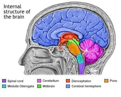
The brain and spinal cord together comprise what is known as the central nervous system (CNS). Like all tumors in the body, CNS tumors are either benign or malignant. Benign tumors are called non-cancerous because they have precise borders, are not invasive, and the cells that make up the growth are similar to other normal cells and grow relatively slowly. However, benign CNS tumors can press on a specific region of the spinal cord or brain and, thereby, cause symptoms. However, when such a benign tumor develops in an area that interferes with essential nervous system functioning, it is treated as malignant.
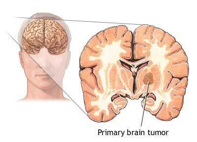
Malignant, or cancerous, tumors of the central nervous system are likely to be fast-growing, are invasive into surrounding healthy tissue, and the cells are very different from normal cells. These tumors can create a life-threatening situation by stopping vital functions of the brain. Some cancerous CNS tumors do not put out roots nor do they grow rapidly. These tumors are described as being encapsulated. Another way that brain and central nervous system tumors are classified is by site of origin. Those that actually develop in the brain or spinal cord are called primary CNS tumors. Metastasis to the brain or spinal cord is, for the most part, a one-way street, meaning these tumors almost never metastasize to other areas in the body. The tumors that develop elsewhere in the body and metastasize, or spread, to the central nervous system are considered to be secondary CNS tumors. Such metastatic cells do not resemble other CNS cells. Instead, they have the same appearance as the cancer cells at the original cancer site elsewhere in the body.
Frequently observed signs of a brain tumor are the following:
Brain and Central nervous system tumors
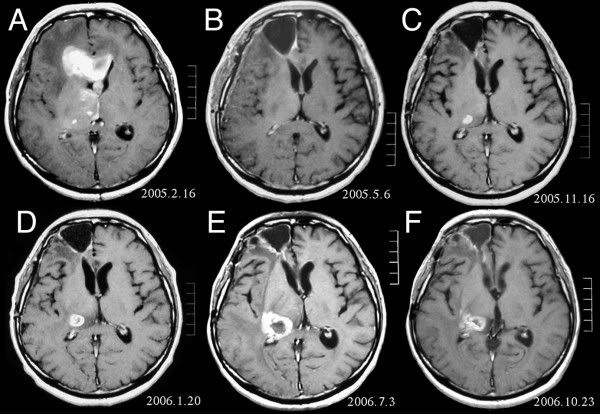
Typically, diagnosis of CNS tumor is made by a physician who does a complete physical examination, including a family history and neurological examination. Computerized tomography (CT) scans, magnetic resonance imaging (MRI) scans, skull x rays, brain scans, angiograms, or myelograms are among the means of visualizing the brain or spinal cord to search for tumors.
General categories of treatment methods for CNS tumors include surgery, radiation therapy, and chemotherapy, with surgery being the single most commonly used therapy. Steroids are usually given prior to treatment to decrease swelling, and anti-convulsant drugs may be given to prevent seizures.
Like all other parts of the body, the brain and central nervous system are made up of cells that ordinarily grow and divide to create new cells as needed. This is usually an orderly process; but when cells lose their ability to grow normally or to die off naturally, they divide too often and produce tumors that are made up of these extra cells.
Description
The brain and spinal cord together comprise what is known as the central nervous system (CNS). Like all tumors in the body, CNS tumors are either benign or malignant. Benign tumors are called non-cancerous because they have precise borders, are not invasive, and the cells that make up the growth are similar to other normal cells and grow relatively slowly. However, benign CNS tumors can press on a specific region of the spinal cord or brain and, thereby, cause symptoms. However, when such a benign tumor develops in an area that interferes with essential nervous system functioning, it is treated as malignant.
Malignant, or cancerous, tumors of the central nervous system are likely to be fast-growing, are invasive into surrounding healthy tissue, and the cells are very different from normal cells. These tumors can create a life-threatening situation by stopping vital functions of the brain. Some cancerous CNS tumors do not put out roots nor do they grow rapidly. These tumors are described as being encapsulated. Another way that brain and central nervous system tumors are classified is by site of origin. Those that actually develop in the brain or spinal cord are called primary CNS tumors. Metastasis to the brain or spinal cord is, for the most part, a one-way street, meaning these tumors almost never metastasize to other areas in the body. The tumors that develop elsewhere in the body and metastasize, or spread, to the central nervous system are considered to be secondary CNS tumors. Such metastatic cells do not resemble other CNS cells. Instead, they have the same appearance as the cancer cells at the original cancer site elsewhere in the body.
Frequently observed signs of a brain tumor are the following:
- severe headaches
- an ataxic, or stumbling, gait
- nausea and vomiting
- lack of coordination
- unusual drowsiness
- weakness or loss of feelings in the arms and legs
- changes in personality or memory
- changes in speech
- changes in vision or abnormal eye movements
- seizures
Brain and Central nervous system tumors
Gliomas
Non-glial tumors
- Astrocytomas
- Brain stem gliomas
- Ependymomas
- Oligodendrogliomas
Non-glial tumors
- Medulloblastomas
- Meningiomas
- Schwannomas
- Craniopharyngiomas
- Germinomas
- Pineal region tumors
General categories of treatment methods for CNS tumors include surgery, radiation therapy, and chemotherapy, with surgery being the single most commonly used therapy. Steroids are usually given prior to treatment to decrease swelling, and anti-convulsant drugs may be given to prevent seizures.


0 comments:
Post a Comment