 Astrocytoma
Definition
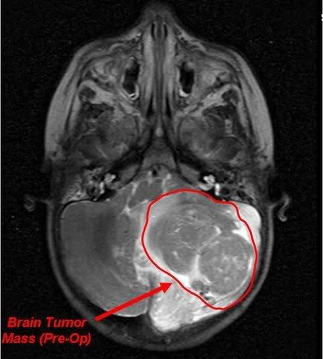
Astrocytoma is a tumor that arises from astrocytes, star-shaped cells that play a supportive role in the brain.
Description
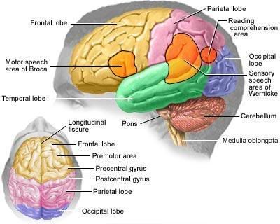
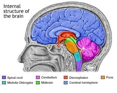
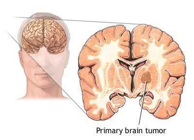
The brain acts as a computer that controls all of the functions of the body. It stores information, memories, and with the use of hormones and electrical impulses, regulates and sends instructions to the rest of the body. Because of the brain’s importance, cancers in the brain can affect many of the body’s functions. The location of a tumor within the brain determines which effects it will have. Astrocytomas may occur in the cerebrum, the site of thought and language, the cerebellum, the area responsible for movement and muscle co-ordination, or the brainstem, the location that regulates critical activities like breathing and heartbeat. Childhood astrocytomas are most commonly located in the cerebellum, while adults usually develop astrocytoma in the cerebrum.
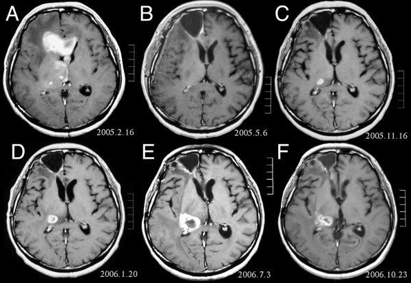
Astrocytomas rarely metastasize (spread) outside the brain to other parts of the body; however, they may grow and spread within the brain. As there is no extra room in the skull, the presence of a brain tumor causes an increase in intracranial (within the skull) pressure, resulting in headaches and possibly affecting normal brain function by compressing delicate brain tissue. Astrocytomas are a type of glioma, a tumor of glial cells (specialized cells that give physical support and electrical insulation between neurons). They are sometimes called gliomas, anaplastic astrocytomas, or glioblastoma multiforme. Oligoastrocytomas are a type of mixed glioma similar to astrocytomas. They usually contain cells that originate from oligodendrocytes as well as astrocytes, and are usually low grade (grading is an estimate of the tumor’s malignancy and aggressiveness; lower-grade tumors require less drastic therapy than high-grade tumors).


0 comments:
Post a Comment