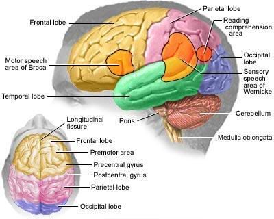
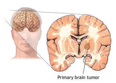
Primary brain tumors are also classified by their site of origin. Gliomas, occurring in the glial, or supportive tissues around the brain, are the most common. Gliomas are further broken down into the following variations:
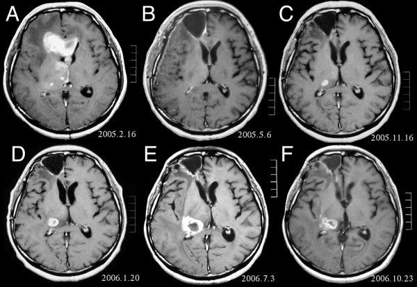
 Fig. 1. Grade Ⅲ craniopharyngioma. T1-weighted coronal-enhanced MR image showing the intracisternal tumor extending to the lower half of the third ventricle.
Fig. 2. A: Grade Ⅳ craniopharyngioma. The patient had acute disturbance of consciousness. Emergency cranial T1-weighted enhanced MR image illustrating that the tumor grew upwards into the upper part of the third ventricle and the right lateral ventricle was slightly dilated. Patient’s condition improved after ventriculoperitoneal shunt. The removed tissues confirmed that the patient had craniopharyngioma. B: The patient well recovered and MRI after 3 years showed no recurrence.
Fig. 3. Grade Ⅴ craniopharyngioma. T1-weighted enhanced magnetic resonance image showing that the tumor manifested itself as an uneven signal and multiform or polycystic form, and the tumor extended into the right lateral ventricle and the brain parenchyma through the interventricular foramen.
Angiogram—A diagnostic test that makes it possible for blood vessels to be seen on film by filling them with a contrast substance or dye that appears on x rays.
Anti-convulsant drugs—A group of medications used in the treatment of seizures.
Brain scan—A general term that can include CT scans, MRIs, seldom-used radionuclide scanning (use of radioactive isotopes), or ultrasounds.
Computerized tomography (CT) scan—The combined use of a computer and x rays that are passed through the body to produce clear, cross-sectional images.
Magnetic resonance imaging (MRI)—An imaging technique that produces good cross-sectional images without x rays or other radiation sources.
Myelogram—X-ray examination of the spinal cord after injection of a contrast substance or dye that shows up on x rays.
Neurological exam—A physical examination that focuses on the patient’s nerves, reflexes, motor and sensory functions, and muscle strength and tone.
Seizures—Sudden, uncontrolled electrical activity in the brain resulting in characteristic twitching, or spastic, movements that may be accompanied by loss of consciousness.
Steroids—A group of drugs that are similar to the hormones produced by the cortex of the adrenal gland.
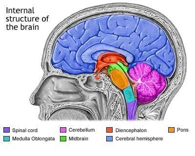
Ventricles of the brain—The four fluid-filled chambers, or cavities, found in the two cerebral hemispheres of the brain, at the center of the brain, and between the brain stem and cerebellum, and linked by channels, or ducts, allowing cerebral fluid to circulate through them.
- Astrocytomas are named for the star-shaped, small cells that they are comprised of. Children may develop these in their brain stem, cerebrum, or cerebellum, while adults commonly develop them in the cerebrum.
- Brain-stem gliomas are usually astrocytomas that originate in the bottom, stem-like portion of the brain. Because this area controls many essential bodily functions, such tumors often cannot be removed.
- Ependymomas occur in the linings of the four brain ventricles, or chambers, or along the spinal cord. These are more common in children.
- Oligodendrogliomas are very rare and, when seen, are usually found in middle-aged adults. They grow slowly and ordinarily do not invade surrounding brain or spinal cord tissue. They originate in the cells responsible for the manufacture of myelin, a fatty covering for nerve tissue.
- Medulloblastomas, tumors of the cerebellum, are most common in male children. Studies have shown these to originate in primitive nerve cells that normally would have disappeared soon after birth.
- Meningiomas are usually benign. They develop in the meninges, or brain linings, and grow very slowly. Because of this slow growth, they may go undetected for years. Meningiomas are more common in women between the ages of 30 and 50.
- Schwannomas are also benign tumors, specific to the myelin-producing cells (Schwann cells) for the acoustic, or hearing, nerve. These, too, are more common in women than men.
- Craniopharyngiomas are usually benign, but because of their location near the pituitary gland and hypothalmus, they can easily affect vital functions and are therefore treated as if malignant. They occur more frequently in children and teenagers.
 Fig. 1. Grade Ⅲ craniopharyngioma. T1-weighted coronal-enhanced MR image showing the intracisternal tumor extending to the lower half of the third ventricle.
Fig. 2. A: Grade Ⅳ craniopharyngioma. The patient had acute disturbance of consciousness. Emergency cranial T1-weighted enhanced MR image illustrating that the tumor grew upwards into the upper part of the third ventricle and the right lateral ventricle was slightly dilated. Patient’s condition improved after ventriculoperitoneal shunt. The removed tissues confirmed that the patient had craniopharyngioma. B: The patient well recovered and MRI after 3 years showed no recurrence.
Fig. 3. Grade Ⅴ craniopharyngioma. T1-weighted enhanced magnetic resonance image showing that the tumor manifested itself as an uneven signal and multiform or polycystic form, and the tumor extended into the right lateral ventricle and the brain parenchyma through the interventricular foramen.
- Germinomas, or germ cell tumors, develop from primitive sex cells called germ cells.
- Pineal-region tumors originate in the area near the pineal gland, a small central brain gland that secretes melatonin, a brain chemical. These can be either fastgrowing pineoblastoma, or slow-growing pineocytoma.
Angiogram—A diagnostic test that makes it possible for blood vessels to be seen on film by filling them with a contrast substance or dye that appears on x rays.
Anti-convulsant drugs—A group of medications used in the treatment of seizures.
Brain scan—A general term that can include CT scans, MRIs, seldom-used radionuclide scanning (use of radioactive isotopes), or ultrasounds.
Computerized tomography (CT) scan—The combined use of a computer and x rays that are passed through the body to produce clear, cross-sectional images.
Magnetic resonance imaging (MRI)—An imaging technique that produces good cross-sectional images without x rays or other radiation sources.
Myelogram—X-ray examination of the spinal cord after injection of a contrast substance or dye that shows up on x rays.
Neurological exam—A physical examination that focuses on the patient’s nerves, reflexes, motor and sensory functions, and muscle strength and tone.
Seizures—Sudden, uncontrolled electrical activity in the brain resulting in characteristic twitching, or spastic, movements that may be accompanied by loss of consciousness.
Steroids—A group of drugs that are similar to the hormones produced by the cortex of the adrenal gland.
Ventricles of the brain—The four fluid-filled chambers, or cavities, found in the two cerebral hemispheres of the brain, at the center of the brain, and between the brain stem and cerebellum, and linked by channels, or ducts, allowing cerebral fluid to circulate through them.


1 comments:
best Dermatologists in hauz khas provide you high level of service.
Post a Comment