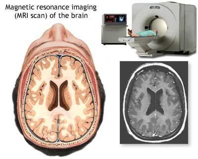
In the first stage of diagnosis the doctor will take a history of symptoms and perform a basic neurological exam, including an eye exam and tests of vision, balance, coordination and mental status. The doctor will then require a computerized tomography (CT) scan and magnetic resonance imaging (MRI) of the patient’s brain. During a CT scan, x rays of the patient’s brain are taken from many different directions; these are combined by a computer, producing a cross-sectional image of the brain. For an MRI, the patient relaxes in a tunnel-like instrument while the brain is subjected to changes of magnetic field. An image is produced based on the behavior of the brain’s water molecules in response to the magnetic fields. A special dye may be injected into a vein before these scans to provide contrast and make tumors easier to identify.
 If a tumor is found it will be necessary for a neurosurgeon to perform a biopsy on it. This simply involves the removal of a small amount of tumor tissue, which is then sent to a neuropathologist for examination and staging. The biopsy may take place before surgical removal of the tumor or the sample may be taken during surgery. Staging of the tumor sample is a method of classification that helps the doctor to determine the severity of the astrocytoma and to decide on the best treatment options. The neuropathologist stages the tumor by looking for atypical cells, the growth of new blood vessels, and for indicators of cell division called mitotic figures.


0 comments:
Post a Comment