Showing posts with label tumors. Show all posts

Brain Tumor : Defination and Description

Astrocytoma


Definition

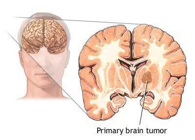
Astrocytoma is a tumor that arises from astrocytes, star-shaped cells that play a supportive role in the brain.

Description
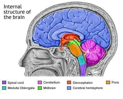
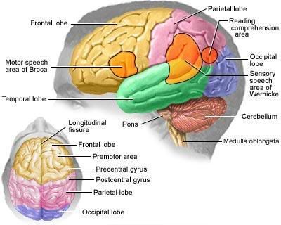
The brain acts as a computer that controls all of the functions of the body. It stores information, memories, and with the use of hormones and electrical impulses, regulates and sends instructions to the rest of the body. Because of the brain’s importance, cancers in the brain can affect many of the body’s functions. The location of a tumor within the brain determines which effects it will have. Astrocytomas may occur in the cerebrum, the site of thought and language, the cerebellum, the area responsible for movement and muscle co-ordination, or the brainstem, the location that regulates critical activities like breathing and heartbeat. Childhood astrocytomas are most commonly located in the cerebellum, while adults usually develop astrocytoma in the cerebrum.

Astrocytomas rarely metastasize (spread) outside the brain to other parts of the body; however, they may grow and spread within the brain. As there is no extra room in the skull, the presence of a brain tumor causes an increase in intracranial (within the skull) pressure, resulting in headaches and possibly affecting normal brain function by compressing delicate brain tissue. Astrocytomas are a type of glioma, a tumor of glial cells (specialized cells that give physical support and electrical insulation between neurons). They are sometimes called gliomas, anaplastic astrocytomas, or glioblastoma multiforme. Oligoastrocytomas are a type of mixed glioma similar to astrocytomas. They usually contain cells that originate from oligodendrocytes as well as astrocytes, and are usually low grade (grading is an estimate of the tumor’s malignancy and aggressiveness; lower-grade tumors require less drastic therapy than high-grade tumors).
QUESTIONS TO ASK THE DOCTOR

  1. Where inside my brain is the cancer located and where will it spread?
  2. What types of treatment are recommended?
  3. What are the possible side effects of this treatment?
  4. How can the side effects be minimized?
  5. Am I eligible for any clinical trials?
  6. Are there any alternatives to this treatment?
  7. What are the chances that the cancer will return?
  8. Will this cause any disabilities?
  9. How will this affect my daily life?
Tumor biopsies may show large areas of necrosis, or dead cells, surrounded by areas of rampant growth. There may also be a mixture of cell types within the biopsy. Genetic studies show that a number of different types of mutations can take place in genes for tumor suppressor p53 and other proteins that play a role in controlling the normal growth of cells.

Often GBM cannot be entirely surgically removed because it affects large areas of the brain. Radiation therapy will be given regardless of whether surgery is possible, except to very young children. Conventional radiation may be performed, but more specialized types, such as stereotactic radiosurgery, which uses imaging and a computer to treat the tumor very precisely, or interstitial radiation, which delivers radiation by placing radioactive material directly on the tumor, may also be used. Chemotherapy will follow radiation; it may include carmustine, lomustine, procarbazine, and vincristine.

GBM is most common in patients over 50 years of age and rarely occurs in patients under 30. Increasing age is associated with a poorer prognosis. Median survivalis 9 to 11 months following treatment. Fewer than 5% of patients are alive five years later. Because of the poor prognosis of GBM, it is treated more aggressively than low-grade astrocytomas; many clinical trials take place to test new treatments.

Alternative and complementary therapies
While no specific alternative therapies have become popular for this particular type of brain cancer, patients interested in pursuing complementary therapies should discuss the idea with their doctor. A doctor may be able to provide information about the efficacy of certain techniques and whether they may interfere with conventional treatment.

Prevention

Currently, scientists do not know what causes the majority of brain cancers. There may be a slight genetic predisposition, as family members of astrocytoma patients have a slightly increased incidence of the disease.

Clinical studies show that a large number of genetic alterations take place in the higher grade astrocytomas; although this helps to explain what is going wrong in the cells, it does not explain what is causing these genetic mutations to take place.

While it is known that ionizing radiation can cause brain tumors, most people are not exposed to this type of radiation unless they are being treated for cancer. Ongoing studies are examining the long-term risks of other types of radiation, but as of 2001, neither x rays, electromagnetic fields, or cellular phones appear to increase the likelihood of brain cancers. Although evidence is not yet conclusive, some studies suggest that some brain tumors may be caused by environmental exposure to certain organic chemicals.

Exposure is most harmful to the developing fetus and infants, so pregnant women may wish to consider whether they have any occupational exposure to organic chemicals. Parents of infants should be aware of pesticides and any other potentially harmful chemical their child could come into contact with.

Additionally there is some evidence that supplements containing vitamins A, C, E, and folate may have a protective effect when taken during pregnancy. The children of women who take these supplements during pregnancy are half as likely to develop brain tumors before age five.

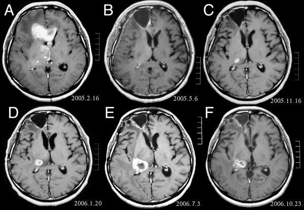
MRI of brain. (A) Initial MRI, shows a tumor in the right and left frontal lobe as well as the right thalamus. (B) MRI after surgery, radiation and chemotherapy. The tumor has completely disappeared except for slight enhancement adjacent to the surgical margin. (C) Recurrence of the thalamic tumor despite maintenance chemotherapy on 9 month after. (D) Increase in size of the thalamic tumor two months after stereotactic radiotherapy. (E) After 6 cycles of TMZ therapy, the thalamic lesion enlarged, and the patient developed dysarthria and hemiparesis. (F) After 2 courses of treatment with interferon-beta and TMZ, the tumor shows a partial response.

Powered by: Blogger | Template and Design by: sifu